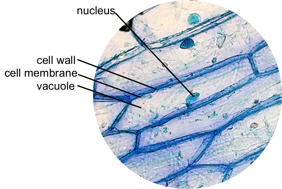
animal cell under light microscope
The seasoned team soon confirmed the death was a murder but no footprints no fingerprints no weapons were founda few strands of hair caught in the dead womans broken fingernails were the only evidence the killer left behind. A nucleus or a cell wall can be seen more clearly by using different stains.

Cells Really Are Like A Kingdom Nikog Animal Cell Cell Diagram Animal Cell Structure
A veterinary pathologist then examines the slide under a microscope.

. Most plant and animal cells are only visible under a light microscope with dimensions between 1 and 100 micrometres. A biopsy is a surgical excision of a piece of the tumor. Make a wet or dry mount with a coverslip.
For example an amoeba is about 1 mm in length and the biggest ones can be seen without a microscope. Much later in 1831 Robert Brown an Englishman observed that all cells had a centrally positioned body which he termed. The cell from the Latin word cellula meaning small room is the basic structural and functional unit of lifeEvery cell consists of a cytoplasm enclosed within a membrane which contains many biomolecules such as proteins and nucleic acids.
Dye is used to stain the cells making them easier to see. Hooke discovered a multitude of tiny pores that he named cells. Although this is not the case with all it is the most common.
One observation was from very thin slices of bottle cork. Pieces of the tumor are then examined by a veterinary pathologist under the microscope. It comprises other cellular structures and organelles which helps in carrying out some specific functions required for the proper functioning of the cell.
In some cases results from FNA may not be entirely clear and biopsy may be necessary. In biological terms an animal cell is a typical eukaryotic cell with a membrane-bound nucleus with DNA present inside the nucleus. When viewed under the light microscope Euglena appear as elongated unicellular organisms that are rapidly moving across the field surface.
Get Forensic with Hair Analysis Life was long gone from the cold bloody corpse when the crime scene investigators arrived. Cells can range in size. Microscope cell staining is a technique used to improve the visibility of cells and cell parts under a microscope.
In 1672 Leeuwenhoek observed bacteria sperms and red blood corpuscles all of which were cells. One thing that students will notice as soon as they begin to observe the organism is that it has a blunt rounded end portion and a pointed end this gives them a tear-drop shape. Iodine crystal violet and methylene blue are examples of simple stains.
1665 observed a piece of cork under the microscope and found it to be made of small compartments which he called cells Latin cell small room. Even though plant cells are eukaryotic the difference can be easily identified as the animal cells lack. Cells can be seen with a light microscope which can magnify objects up to 1000 times.
Typically a microscope slide is prepared which creates a thin layer of cells and holds them in place. The cell was first discovered by Robert Hooke in 1665 which can be found to be described in his book Micrographia. In this book he gave 60 observations in detail of various objects under a coarse compound microscope.
This is called histopathology.

Cell 8 Pictures Of Plant Cells Under A Microscope Plant Cell Structure Under Microscope Plant And Animal Cells Plant Cell Structure Plant Cell Picture

Animal Cell Structure Cell Organelles Organelles

Epidermal Onion Cells Under A Microscope Plant Cells Appear Polygonal From The Cell Diagram Plant Cell Diagram Plant Cell

Year 11 Bio Key Points Cell Organelles And Their Function Cell Organelles Animal Cell Organelles

Histolab4a Htm Histology Slides Science And Nature Animal Cell

Animal Cell Coloring Printing 5 Animal Cell Drawing In Cell Category Human Cell Diagram Cell Diagram Animal Cell Drawing

Animal Cell Organelles Sauna Design

Cheek Cells Things Under A Microscope Cell Cheek

Structure Of Animal Cell And Plant Cell Under Microscope Diagrams Cell Diagram Plant Cell Diagram Animal Cell

An Electron Micrograph Of A Mouse Liver Cell Dna Learning Center Electrons Cell Learning Centers

Animal And Plant Cell Microscope Slide Set Set Of 2 Frey Scientific Symmetry In Microscopy Plant Plant And Animal Cells Animal Cell Plant Cell Picture

Plant And Animal Cells Revised Plant Cell Project Animal Cell Project Plant And Animal Cells

Onion Cells Under A Microscope Requirements Preparation Observation Plant And Animal Cells Animal Cell Plant Cell

Editible Eps Vector File The Animal Cell Diagram Vector Etsy Animal Cell Cell Diagram Plant Cell Diagram

560 X 364 Pixel Electron Microscope Image Animal Cell And Organelles Labeled Animal Cell Plasma Membrane Organelles

Cell Theory Plant Cell Diagram Cell Diagram

Microscopic Animal Cells Images Kuhn Photo Microscopic Cells Microscopic Photography Things Under A Microscope

Onion Epidermis Under Light Microscope Purple Colored Large Microscopic Photography Microscopic Microscope
